
Page 21 - An Innovation Spirit ...
P. 21
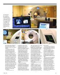
Twin state-of-the-
art CT scanners
have allowed the
Peter Munk Cardiac
Centre to “rapidly put
cutting-edge research
protocols into clinical
care as soon as they
are validated,” says
Dr. Narinder Paul.
He calls it a “huge”
advancement in the
study and care of
patients with heart
disease.

partnership with the JDMI and          pushing the envelope of how          able to give much greater accuracy    Mr. Dettmann.
the centre that aims to make           quickly we are able to capture       and speed, so you can assess           But the company is also looking
imaging procedures more “patient       an anatomic picture of the           things now you absolutely couldn’t
friendly.”                             human body, and that’s of critical   do five years ago,” says Dr. White.   at preparing for future health-care
                                       importance in cardiac imaging.                                             needs, and that’s where important
 This is what a patient with a         Imagine the heart as a piece           Dr. Paul adds: “With a cardiac      medical partnerships –
possible heart condition can           of muscle beating and moving         CT, because patients may be           such as the one with the Joint
expect to experience as part of        rapidly in three dimensions,         nervous, the heart rate can be        Department of Medical Imaging
the joint CT imaging project:          and the inherent challenges in       quite fast. Therefore, we often       and the Peter Munk Cardiac
The patient undergoes a CT scan,       capturing anatomic details of this   have to temporarily slow the heart    Centre – come into play, he says,
during which the dye injected          complex anatomic structure in a      down to 60 beats per minute           adding that there currently are
into a vein will go to wherever the    single snapshot in time,” says Dr.   using medication. With these          no plans to put this research-to-
blood goes – if there’s a restriction  Lawrence White, Radiologist-in-      new machines, because the X-ray       clinical platform anywhere but the
to blood going into a certain area,    Chief of the Joint Department of     tube and detector spin that much      PMCC.
that dye will not highlight as well    Medical Imaging, a joint program     faster, the heart rate can be as
as it would in areas where there       of radiology at Mount Sinai          high as 80 beats per minute, and       “What you need is the physicians
is no restriction. The image data      Hospital, the University Health      we can still obtain wonderful         telling you: what the headaches
then goes to a powerful computer       Network and Women’s College          images at a fraction of the           are you have now; what the
and a software program, where          Hospital.                            radiation dose that is used in older  solutions are you’re looking for;
the heart can be turned in three                                            CT scan units.”                       what things we need to do in
dimensions to look at every vessel,      “When trying to assess                                                   five to 10 years; and what kinds
no matter how small – both in and      something as small as the              So where does the future lie for    of diseases are increasing,” Mr.
around the heart. This determines      coronary arteries, the vessels that  imaging?                              Dettmann says. “Definitely, heart
how blood is flowing into that         supply blood to the heart muscle,                                          disease is one of the top two
area, thus seeing if there’s any       to be able to see and assess these    Toshiba of Canada, for one, is       diseases in the world [along with
disease, the extent of the disease     structures you need a certain        focusing on “volume” scanners –       cancer], so having a solution to
and whether it requires any            amount of resolution to your         ones that capture 3-D and even        the variety of heart diseases, and
intervention.                          images; they need to be sharp and    4-D images that cover more area       developing them together with
                                       accurate. These new machines are     and in less time than conventional    clinicians, is a very important part
  “These new scanners are                                                   scanners, meaning less radiation      of our business.”
                                                                            exposure and quicker results, says

Winter 2016                                                                                                                                             19