
Page 27 - An Innovation Spirit ...
P. 27
Graham Nie sat down better-than-fighting chance when for the patient, says Dr. Tan, because
with his doctor at the a large aneurysm caused a tear in X-ray dye can be toxic to the kidneys.
Peter Munk Cardiac his aorta. In February 2013, doctors
Centre (PMCC) to look at at the PMCC implanted a stent (an With the PMCC’s advanced
a CT scan of his heart, expandable tube made with fabric imaging technology, a CT scan is
rendered in 3-D on a and alloy materials) into the affected used to generate a 3-D picture of the
computer screen, a year artery using a tube inserted through a aorta, which is then fused with an
after he had surgery to small slit in the groin. Once the stent X-ray of the same area. The result
implant a stent in a was in place, a balloon tip at one end is a complete and detailed digital
diseased artery. of the tube inflated it to its full size, representation of the artery, allowing
enabling it to reinforce the weakened for precise placement of the stent
“It was amazing. He could flip this artery and prevent further swelling. without further injections of X-ray
image around, so I could see where dye.
the stent and its branches were put “Advanced EVAR is one of the most
in,” recalls Mr. Nie, who was first significant developments in aortic “What is unique here is our ability to
diagnosed in 1997 with an abdominal surgery, developed elsewhere, but offer advanced EVAR in an advanced
aortic aneurysm – a balloon-like bulge refined at UHN,” says Dr. Kong Teng imaging environment,” says Dr.
that can cause parts of the body’s Tan, Division Head, Interventional Tan, referring to the image-guided
largest blood vessel, the aorta, to swell Radiology, Toronto General Hospital. operating rooms (OR), which feature
and, in some cases, rupture. “It was large computer screens that give OR
the first time in many years that I was “The conventional way is open teams access to CT, X-ray and other
clear of aneurysms.” surgery, with an incision practically images during a surgical procedure.
from the top of the chest to the groin,” “We are one of the few pioneering
Today, Mr. Nie, a 75-year-old retired adds Dr. Tan, noting that the PMCC places to do it.”
school principal, remains aneurysm- leads the country in the number of
free and continues to recover. He’s advanced EVARs performed each The use of CT scans in cardiology is,
active, albeit slower in his everyday year. “It’s very invasive, so not all in itself, a pioneering strategy that the
tasks such as mowing the lawn around patients can go through the operation PMCC continues to advance through
his property in Peterborough, Ont. because you have to have a certain research. Aside from minimizing
level of health to tolerate it.” X-ray dye injections, CT scans provide
Mr. Nie’s successful treatment and deeper insight into the anatomy of
recovery can be attributed directly With advanced EVAR, the PMCC the heart, all the way down to the
to leading-edge innovations at the can give more patients a chance to cellular level. This allows doctors to
PMCC, particularly in advanced get a potentially life-saving stent – an see physiological clues and diagnose
medical imaging. From the increased avenue that used to be closed off to disease sooner and more precisely.
use of CT scans to diagnose and about 80 per cent of patients with For example, the amount of blood
address heart and vascular disease aortic aneurysms, says Dr. Tan. But flow to muscles in the heart might
to multimodal practitioners with what sets the PMCC apart from many indicate that certain arteries are
combined expertise in cardiology, other hospitals that offer advanced likely to continue narrowing and will
vascular surgery and radiology, the EVAR is its use of advanced imaging eventually stop functioning.
PMCC is driving advances in medical during the procedure.
methods and technology to improve “By looking at the physiology, we can
patient outcomes. In the past, doctors had to keep pick up disease early and characterize
injecting patients undergoing EVAR the disease to help with decision
Located at Toronto General with X-ray dye to ensure the stent was making,” says Dr. Paul. “This type of
Hospital – one of four hospitals that positioned properly. This isn't good insight also helps us determine later if
make up Ontario’s University Health the treatment is working.”
Network (UHN) – the PMCC has a
history of innovations that goes back Advanced uses and research with
75 years. It boasts many world firsts,
from the first pacemaker implant in CT scans aren’t the only imaging
1950 to the first installation and use
of the Carto 3 heart monitor recorder innovations at the PMCC. Dr. Danna
system, which uses electromagnetic
technology to create three- Spears, Clinical Director of the
dimensional images of the heart.
Heritable Arrhythmia Program at
“Everything we do is based around
how to make things better for patients the PMCC, uses catheter-guiding
and for the health-care system,” says
Dr. Narinder Paul, Division Chief, technologies to map patients’ cardiac
Cardiothoracic Radiology, University
Health Network. The medical imaging electrical systems by picking up
team that also serves Toronto’s Mount
Sinai and Women’s College hospitals. signals from an
“This means exploring and embracing
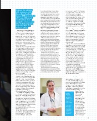
new technologies, as well as Dr. Kong ultrasound probe
innovating through new applications Teng Tan or by sensing
of existing technology.” and Dr. Elsie changes in
Nguyen are electrical resistance.
In Mr. Nie’s case, the marriage of two members of Electrode patches
innovations – advanced endovascular the innovative applied on the
aneurysm repair (EVAR) and
advanced imaging – gave him a medical body generate a
imaging team. magnetic field that
The work of communicates
Dr. Danna with an electrode
Spears (near tip on the catheter
left) focuses to produce 3-D
on patients images of the heart’s
with irregular electrical system.
heartbeats.
“The conventional
way of imaging is
25